Chest X-Ray: Reasons for Procedure, Normal and Abnormal Results

Welcome to the world of chest X-rays—a cornerstone of medical imaging that lets healthcare professionals peer inside your body with incredible detail and speed. Whether you're dealing with a persistent cough, chest pain, or simply undergoing a routine check-up, a chest X-ray provides vital insights that guide diagnosis and treatment.
From detecting infections like pneumonia to identifying chronic conditions like emphysema, chest X-rays are invaluable tools in modern medicine. In this article, we’ll explore everything you need to know about chest X-rays: how they're performed, what you can expect during the procedure, and the benefits and risks involved. Let's dive in!
Chest X-rays are one of the most common diagnostic tools used in medicine today. These imaging tests are quick, non-invasive, and can provide a wealth of information about the health of your lungs, heart, and chest wall. When you get a chest X-ray, a small amount of ionising radiation is used to produce images of the inside of your chest.
Chest X-rays involve standing or sitting in front of a specialised plate that captures the image as the X-ray passes through your body. It's a painless and swift process, generally completed within a few minutes. Radiographers will position you correctly and make sure to use radiation protection devices to minimise your exposure.
Overall, chest X-rays are a valuable tool in modern medicine, enabling your healthcare provider to get a clear picture of your chest's internal structures. In the following sections, we'll delve deeper into how chest X-rays work, what you can expect during the procedure, and how to interpret the results.
What is a chest X-ray?
A chest X-ray is a type of medical radiography specifically aimed at capturing detailed images of your chest area. This procedure uses X-rays, which are a form of electromagnetic radiation, to penetrate your body and create pictures of the internal structures. By revealing the state of your heart, lungs, airways, blood vessels, and bones of your spine and chest, it provides crucial information to your healthcare provider.
How does it work?
During a chest X-ray, you will usually be asked to stand against a flat surface while the radiographer captures the images. The X-rays pass through your body and onto a film or a digital detector. Different tissues absorb varying amounts of radiation, which is why bones appear white, air appears black, and soft tissues appear in shades of gray on the resulting images.
Why might you need one?
Chest X-rays are incredibly useful for diagnosing various conditions such as pneumonia, heart issues, lung cancer, and other lung problems. If you are experiencing symptoms like persistent cough, chest pain, or shortness of breath, your doctor may recommend a chest X-ray to help pinpoint the cause and formulate an effective treatment plan.
Understanding what a chest X-ray is and how it works can help alleviate any apprehensions you might have about the procedure. It’s a quick, non-invasive test that plays a vital role in modern medical diagnostics.
How do I prepare for a chest X-ray?
Preparing for a chest X-ray is relatively straightforward, but there are some essential steps you need to follow to ensure the procedure goes smoothly. Here’s what you need to know:
- Wear the Right Clothing: You might be asked to change into a hospital gown to prevent any interference from clothes or metal objects. Loose, comfortable clothing and minimal jewellery are recommended.
- Remove Metal Objects: Metal can obstruct the X-ray image and lead to inaccurate results. Make sure to remove any necklaces, earrings, piercings, or metal hairpins before the procedure.
- Follow Specific Instructions: Your healthcare provider will inform you if there's anything specific you need to do before the X-ray. This may include avoiding food or drinks for a certain period if additional tests are involved.
During the procedure, the radiographer will guide you through each step to ensure accurate images are captured. Don’t hesitate to ask questions if any instructions are unclear. Remember, your comfort and safety are a top priority!
How is the procedure performed?
The chest X-ray procedure is relatively quick and non-invasive, making it a commonly used diagnostic tool. Here's a step-by-step guide on what you can expect:
Positioning
Once you're ready, the radiographer will help you position your body before the X-ray machine. Typically, you will be asked to stand or sit while your chest is pressed against a flat surface called a detector. In some cases, you might be asked to lie down. Proper positioning ensures that the X-ray captures clear images of your heart, lungs, and chest wall.
Taking the X-ray
When you're in the correct position, the radiographer will step behind a protective screen or leave the room to avoid radiation exposure. They will then instruct you to hold your breath briefly while the X-ray is taken. This minimises movement and helps produce a sharp image.
Multiple Views
Several images may be taken from different angles to get a comprehensive view of your chest. This often includes a front (posterior-anterior) view and a side (lateral) view. The radiographer will assist you in repositioning between each shot.
Post-Procedure
After the X-rays are taken, you can usually resume your normal activities immediately. The images are then reviewed by a radiologist who analyses them for any signs of abnormalities or conditions. Your doctor will discuss the results with you and recommend any further tests or treatments if necessary.
Overall, a chest X-ray is a simple yet powerful tool that plays a crucial role in diagnosing many medical conditions affecting the chest and lungs. By understanding the procedure, you can approach it with confidence and ease.
What happens after a chest X-ray?
If the X-ray reveals any abnormalities, further testing might be required. This could include additional imaging studies like a CT scan, MRI, or even a biopsy, depending on what was identified in the X-ray. Your doctor will guide you through these procedures if they become necessary.
When it comes to understanding the benefits and risks of chest X-rays, it’s essential to weigh them carefully, especially since this diagnostic tool plays a crucial role in modern medicine.
Benefits
- Quick and Painless: One of the primary advantages of chest X-rays is the speed and ease with which they can be performed. The entire process is quick, often taking just a few minutes, and is generally painless for the patient.
- Diagnostic Accuracy: Chest X-rays are highly effective for diagnosing a range of conditions such as pneumonia, lung cancer, and heart failure. They provide clear images that help physicians to make accurate diagnoses and treatment plans.
- Non-Invasive: Unlike surgical procedures or other more invasive diagnostic techniques, chest X-rays are non-invasive. This reduces recovery time and the potential for complications.
- Cost-Effective: In comparison to more advanced imaging techniques like MRI or CT scans, chest X-rays tend to be more affordable, making them accessible to a larger population.
Risks
- Radiation Exposure: The primary risk associated with chest X-rays is exposure to ionising radiation. While the dose is generally low, repeated exposure can accumulate over time, potentially increasing the risk of developing radiation-related conditions.
- Over-Reliance: There's a risk of over-relying on chest X-rays when additional diagnostic tests might be necessary. A clear X-ray does not always mean the absence of disease, and conversely, some abnormalities may require further investigation through other imaging techniques.
- Pediatric Concerns: For children, there is an additional concern regarding their sensitivity to radiation.
- Children and pregnant women are particularly vulnerable. In children, growing tissues are more sensitive to radiation. For pregnant women, there's a risk, albeit small, of harming the developing fetus. Radiographers take extra precautions, such as using lead aprons, to minimise exposure in these cases.
The key is to engage in an informed dialogue with your healthcare provider. They can help you understand when the benefits of a chest X-ray outweigh the risks, ensuring you receive the best possible care.
What are the complications associated with a chest X-ray?
Radiation Exposure: One of the main concerns with any X-ray procedure is radiation exposure. Although a chest X-ray involves a relatively low dose, repeated exposure over time can accumulate and potentially lead to adverse effects, including an increased risk of cancer. Radiographers use protective measures like lead shielding to minimise your exposure.
Discomfort or Pain: While not common, you might experience some discomfort or pain during a chest X-ray. This can stem from positioning, especially if you have a pre-existing condition that makes movement painful. However, any discomfort is usually brief and subsides quickly.
Allergic Reactions: In rare cases, patients may have an allergic reaction to the materials used in protective equipment or the environment of the imaging room. It's always wise to inform your healthcare provider of any known allergies beforehand.
False Positives or Negatives: A potential complication is the risk of diagnostic errors, such as false positives or negatives. This can lead to unnecessary stress, further testing, or even incorrect treatment. It's essential to have X-rays interpreted by experienced radiologists to reduce these risks.
Despite these potential complications, chest X-rays are invaluable for diagnosing conditions such as pneumonia, tuberculosis, and lung cancer. The benefits often far outweigh the potential risks, particularly when protective measures are in place. Always discuss any concerns with your healthcare provider to ensure you are fully informed and comfortable with the procedure.
The interpretation of chest X-rays is conducted by specialised medical professionals called radiologists. Radiologists are doctors who are trained to read and analyse medical images, including X-rays, MRI, and CT scans. They employ their expertise to identify any abnormalities or issues within your chest, such as infections, fractures, or tumours.
Once your chest X-ray has been taken, it will be sent to the radiologist. The radiologist closely examines the images and looks for signs that might explain your symptoms or help in diagnosing any medical conditions. Following a thorough review, they will compile their findings into a detailed report.
The time it takes to get the results can vary. In urgent cases, such as potential pneumonia or a suspected fracture, a preliminary interpretation might be available within hours. For routine check-ups, you might receive your results within a few days. Your healthcare provider will then review the radiologist's report and discuss it with you at your next appointment, or they may contact you sooner if immediate action is needed.
If you have any questions or need further explanations about your X-ray results, don't hesitate to ask your healthcare provider. Understanding your results fully can help you make informed decisions about your health and any necessary treatments.
Why might I need a chest X-ray?
Chest X-rays are a powerful diagnostic tool used by healthcare professionals for a variety of reasons. Here are some common scenarios where a chest X-ray might be essential:
- Detecting Lung Conditions: Whether it's pneumonia, tuberculosis, lung cancer, or chronic obstructive pulmonary disease (COPD), chest X-rays can reveal abnormalities in your lungs.
- Heart Problems: Your doctor may order a chest X-ray to check for heart conditions such as heart failure, as it shows the shape and size of the heart and any related issues in the surrounding blood vessels.
- Monitoring Medical Treatments: If you're undergoing treatment for a lung or heart condition, regular chest X-rays may be necessary to monitor your progress and the efficacy of the treatment.
- Injury or Trauma: After an accident, a chest X-ray can help identify fractures, rib injuries, or damage to the lungs.
- Pre-surgical Assessment: Before certain surgeries, doctors often require a chest X-ray to ensure there are no underlying conditions that might complicate the procedure.
- Symptoms Investigation: If you're experiencing unexplained symptoms like persistent cough, shortness of breath, chest pain, or fever, a chest X-ray can help pinpoint the cause.
- Occupational Health: For individuals exposed to dust, chemicals, or other hazards in certain workplaces, routine chest X-rays can be part of a preventive health check.
In all these scenarios, the ultimate goal is to provide a clear and comprehensive picture that aids in accurate diagnosis and effective treatment planning.
How do doctors interpret a chest X-ray?
Interpreting a chest X-ray is a meticulous process undertaken by skilled radiologists. First, they evaluate the overall quality of the X-ray image to ensure it was taken correctly. This involves checking for proper positioning, adequate exposure, and clarity.
Next, radiologists systematically analyse the X-ray, often using a structured approach known as the ABCDE method:
- Airways: They look at the trachea and major bronchi to ensure they are clear and properly aligned.
- Bones: They examine the ribs, clavicles, and spine for any signs of fractures or abnormalities.
- Cardiac: They assess the size and shape of the heart, looking for signs of enlargement or abnormal contours.
- Diaphragm: They check the diaphragm for its position and contour, ensuring it appears normal.
- Extra: Finally, they review additional structures such as tubes, lines, and soft tissues for any unusual findings.
This systematic evaluation helps radiologists uncover issues such as infections, tumours, lung conditions, heart disease, and more.
Once they have thoroughly examined the X-ray, radiologists compile a detailed report. This report is then shared with the referring physician, who will discuss the findings with you and outline any necessary next steps.
Interpreting a chest X-ray requires both expertise and attention to detail. By analysing these images, doctors can provide critical insights into your health, aiding in accurate diagnosis and effective treatment planning.
What is a shadow on a chest X-ray?
When you undergo a chest X-ray, the resulting image may show various "shadows" that can indicate different aspects of your health. But what exactly are these shadows?
A shadow on a chest X-ray is an area where the X-ray beam is absorbed or deflected more than surrounding tissues. This phenomenon occurs because different tissues and structures within your chest cavity absorb X-rays to varying degrees, appearing as various shades of black and white on the film or computer screen.
For instance, air-filled lungs will appear darker because they allow more X-rays to pass through. Conversely, denser structures like bones, hearts, and tumours will appear lighter. These lighter regions are what radiologists refer to as "shadows."
Deciphering these shadows enables doctors to diagnose a range of conditions, from lung infections and fractures to more serious ailments like tumours and heart issues. Knowing what each shadow represents helps in creating a targeted treatment plan tailored to your specific medical needs.
Fluid Buildup: Another telltale sign of CHF that can be observed in an X-ray is the presence of fluid in the lungs. This condition, known as pulmonary oedema, often manifests as white or cloudy areas on the X-ray, indicating fluid accumulation.
Other Indicators: Besides the enlargement of the heart and fluid buildup, chest X-rays can also reveal changes in the blood vessels and in certain patterns of lung markings. These changes are crucial for diagnosing CHF and understanding its severity.
What are the chest X-ray findings for heart failure?
Chest X-rays are a crucial tool in evaluating heart failure. They provide a clear image of the chest's internal structures, including the heart, lungs, and blood vessels. Here are some key findings that radiologists look for:
- Cardiomegaly: An enlarged heart, known as cardiomegaly, is often a primary sign of heart failure. This finding helps clinicians understand the extent to which the heart is overworking.
- Pulmonary Congestion: Fluid accumulation in the lungs, or pulmonary congestion, appears as enhanced markings in the lung fields. This indicates elevated pressure in the pulmonary veins, a common feature of heart failure.
- Pleural Effusion: Excess fluid between the layers of the pleura surrounding the lungs is another sign. Pleural effusion can be seen as a white area on the X-ray, often at the lung bases.
- Kerley B Lines: These are thin, horizontal lines seen on the outer regions of the lungs. They suggest interstitial fluid buildup and are specific markers of pulmonary oedema associated with heart failure.
- Upper Lobe Diversion: Blood is redirected to the upper lobes of the lungs due to increased pulmonary pressure. This upper lobe diversion indicates advanced heart failure and is visible as prominent veins in the upper lung zones.
When should I call my doctor?
After undergoing a chest X-ray, it's crucial to stay vigilant about your health. While most procedures are routine and uneventful, there are times when you should seek medical advice.
If you experience any severe pain or discomfort that persists beyond a few hours, it's a good idea to contact your doctor. This could be a sign of an underlying issue that needs prompt attention.
Notice any unusual symptoms such as difficulty breathing or persistent coughing? Don't hesitate to reach out. These may indicate a complication or an issue that wasn't fully revealed by the X-ray.
Additionally, if you develop redness, swelling, or a rash at the site where the X-ray was performed, it's better to be safe than sorry and get in touch with your healthcare provider.
Remember, your radiologist and doctor are there to help you interpret the results and guide you through the next steps in your healthcare journey. Better to call with concerns than to overlook potential issues.
How many images are taken in a chest X-ray (CXR)?
In a standard chest X-ray (CXR), typically two images are taken. The rationale behind this lies in the need to capture comprehensive and accurate details of the chest area, including the lungs, heart, and bones. These images are usually taken from different angles.
Firstly, there's the Posteroanterior (PA) view. In this view, you stand facing the X-ray film, with the X-ray machine positioned behind you. This helps to give a clear picture of the lungs, as the X-ray beam passes from your back to the front. It’s often considered the most crucial image in a chest X-ray examination, as it provides the best detail with minimal heart magnification.
The second image is the Lateral view. For this image, you stand with one side of your chest against the X-ray film, and your arms raised above your head. This side view helps to pinpoint abnormalities that might be hidden in the PA view and offers a different perspective on the chest structures.
In certain situations, more images may be required. For instance, if an abnormality is detected and needs further investigation, or if the patient is unable to stand and standard views are not possible, additional images might be necessary. These can include oblique or specialised views tailored to provide a thorough assessment.
In summary, chest X-rays serve as a fundamental tool in medical diagnostics, providing crucial insights into the condition of your lungs and heart. By understanding the process—from preparation and positioning to the actual taking of the X-ray and the multiple views captured—you are better equipped to appreciate the benefits and weigh the risks associated with this common procedure. With advances in technology and continuous research, chest X-rays remain an invaluable asset in diagnosing and monitoring various health conditions, ensuring you receive timely and accurate medical care.

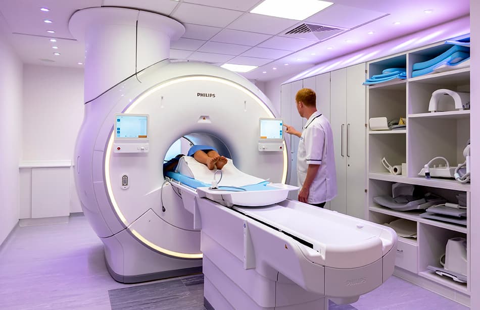
Patient Friendly MRI Scan at UME Health
Our open MRI machine provides a more spacious and less enclosed environment, which can ease any feelings of anxiety or discomfort during the scan. We also allow a friend or family member to sit in the room with you during the scan, if that would make you more comfortable.


