The Marvels of MRI: Discovering the Hidden Secrets of Medical Imaging

In the world of medical imaging, Magnetic Resonance Imaging (MRI) stands as a remarkable innovation that has revolutionised the way we diagnose and understand various health conditions. Fueled by magnetic fields and radio waves, MRI provides a detailed and comprehensive view of the internal structures of the body, allowing doctors to delve deeper into the mysteries of the human anatomy.
With its non-invasive nature, MRI has quickly become a preferred choice for diagnosing and monitoring a wide range of medical conditions, from identifying brain tumours to detecting torn ligaments and even evaluating the health of the heart. This advanced technology offers a wealth of detailed information, enabling healthcare professionals to make accurate and informed decisions when it comes to patient care.
Through this article, we will unlock the marvels of MRI and delve into the hidden secrets it unveils. From its inception to the technology behind it, we will explore the various applications of MRI and its role in diagnosing complex medical conditions. Get ready to dive into the fascinating world of medical imaging and discover how MRI uncovers the hidden secrets of the human body.
How MRI works
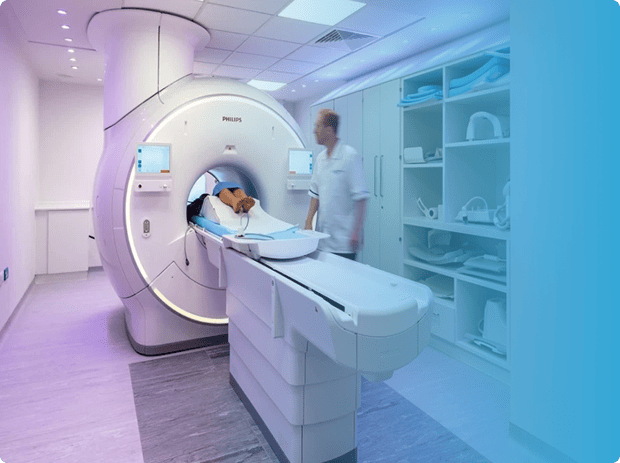
MRI works by utilising a powerful magnetic field and radio waves to create detailed images of the body's internal structures. Unlike X-rays or CT scans, which rely on ionising radiation, MRI is considered safe and non-invasive. The process begins with the patient lying down on a table that slides into the MRI machine. This machine contains a large magnet and radiofrequency coils that work together to generate the images.
When the magnetic field is applied, the protons in the body's atoms align with the field. Radio waves are then used to disturb this alignment, causing the protons to emit energy signals. These signals are detected by the MRI machine and converted into highly detailed images of the body's tissues and organs. The entire process is painless and does not involve any exposure to harmful radiation.
MRI technology has come a long way since its inception. Early MRI machines were large and slow, producing low-resolution images. However, advancements in technology have led to the development of more compact and efficient machines, capable of producing high-resolution images in a fraction of the time. These modern machines also offer a range of imaging techniques, allowing healthcare professionals to tailor the scan to the specific needs of each patient.
Benefits and applications of MRI
The benefits of MRI are numerous and far-reaching. One of the key advantages is its ability to produce detailed images of soft tissues, such as the brain, muscles, and organs. This makes MRI particularly valuable in diagnosing conditions that may not be easily detected by other imaging techniques. For example, MRI can help identify brain tumours, detect spinal cord injuries, and evaluate the health of the heart and blood vessels.
MRI is also a valuable tool for monitoring the progression of certain medical conditions. By conducting regular MRI scans, doctors can assess the effectiveness of treatments, track the growth of tumours, and evaluate the healing process of injuries. This allows for personalised and targeted care, ensuring that patients receive the most appropriate treatments based on their individual needs.
Furthermore, MRI is a non-invasive procedure, meaning that it does not require any surgical incisions or injections. This makes it a safer and more comfortable option for patients, particularly those who may be sensitive to radiation or have a fear of enclosed spaces. Additionally, MRI does not use any harmful contrast dyes, minimising the risk of allergic reactions or side effects.
Types of MRI scans
There are several types of MRI scans that can be performed, each tailored to capture specific information about the body. Some common types include:
- T1-weighted imaging: This type of MRI scan provides detailed anatomical information, highlighting the contrast between different tissues. It is commonly used to evaluate the brain, spine, and joints.
- T2-weighted imaging: T2-weighted imaging is used to visualise fluid-filled structures, such as the cerebrospinal fluid in the brain or the synovial fluid in joints. This type of scan is particularly useful in identifying tumours, infections, and inflammations.
- Diffusion-weighted imaging: Diffusion-weighted imaging is highly sensitive to the movement of water molecules in tissues. It is commonly used to assess strokes, brain injuries, and tumours, as well as to detect abnormalities in organs such as the liver and prostate.
- Functional MRI (fMRI): fMRI is a specialised type of MRI scan that measures changes in blood flow to specific regions of the brain. It is commonly used in neuroscience research to study brain activity and can help identify areas of the brain responsible for certain functions, such as language or movement.
These are just a few examples of the many types of MRI scans available. The specific type of scan recommended will depend on the medical condition being investigated and the information required by the healthcare professional.
MRI vs other medical imaging techniques
When it comes to medical imaging, there are several techniques available, each with its own advantages and limitations. MRI stands out in several aspects, making it a preferred choice for many healthcare professionals.
Compared to X-rays and CT scans, which use ionising radiation, MRI does not expose patients to harmful radiation. This is particularly important for individuals who require multiple scans or those who are more sensitive to radiation, such as children and pregnant women. Additionally, MRI provides better soft tissue contrast, allowing for more detailed visualisation of organs and structures.
Ultrasound, another commonly used imaging technique, relies on sound waves to create images of the body. While ultrasound is effective for certain applications, such as monitoring pregnancy or visualising blood flow, it may not be as useful for evaluating complex anatomical structures. MRI, on the other hand, provides a more comprehensive view of the body, making it an ideal choice for diagnosing a wide range of conditions.
It is worth noting, however, that each imaging technique has its own strengths and limitations. Depending on the specific needs of the patient, healthcare professionals may recommend a combination of imaging techniques to obtain a complete and accurate diagnosis.
Preparing for an MRI scan
If you have been scheduled for an MRI scan, there are a few steps you can take to ensure that the procedure goes smoothly. It is important to follow any instructions provided by your healthcare provider, as these may vary depending on the type of scan being performed.
Firstly, it is important to inform your healthcare provider about any metal implants or devices in your body. Certain metallic objects, such as pacemakers, implants, and even certain tattoos, can interfere with the magnetic field used in MRI. In some cases, these objects may need to be removed or additional precautions may need to be taken.
You may also be asked to refrain from eating or drinking for a specific period of time before the scan, particularly if contrast dye will be used. Additionally, it is important to let your healthcare provider know if you have any allergies or have had adverse reactions to contrast dyes in the past.
It is also common for patients to be asked to change into a hospital gown before the scan, as certain types of clothing can interfere with the magnetic field. It is advisable to dress comfortably and avoid wearing any jewellery or accessories that contain metal.
What to expect during an MRI scan
During an MRI scan, you will be asked to lie down on a table that slides into the MRI machine. It is important to remain as still as possible during the scan, as movement can blur the images. You will typically be provided with earplugs or headphones to minimise the noise generated by the machine, as MRI machines can produce loud knocking or buzzing sounds.
The scan itself is painless and usually takes between 30 minutes to an hour, depending on the type of scan being performed and the area of the body being imaged. It is important to communicate with the healthcare professionals conducting the scan if you experience any discomfort or anxiety during the procedure. They may be able to provide you with additional support or take steps to alleviate any discomfort.
Once the scan is complete, a radiologist will review the images and provide a report to your healthcare provider. The results will then be discussed with you during a follow-up appointment, where further treatment options or recommendations may be provided.
Common misconceptions about MRI
Despite its widespread use and proven effectiveness, there are still some misconceptions surrounding MRI. One common misconception is that MRI is always claustrophobic and uncomfortable. While it is true that some individuals may feel anxious or claustrophobic during the scan, modern MRI machines are designed with patient comfort in mind. Open MRI machines, for example, offer a more spacious and less enclosed environment, reducing feelings of claustrophobia.
Another misconception is that MRI scans are always loud and noisy. While it is true that MRI machines can produce loud knocking or buzzing sounds, patients are typically provided with earplugs or headphones to minimise the noise. Additionally, some newer MRI machines have been designed to produce less noise, making the experience more comfortable for patients.
It is also important to dispel the misconception that MRI scans are harmful due to the use of magnetic fields. MRI machines use strong magnetic fields, but they are not known to cause any harm to the body. However, it is important to inform your healthcare provider if you have any metallic implants or devices, as these can interfere with the magnetic field and may require additional precautions.
Advancements in MRI technology
Over the years, MRI technology has undergone significant advancements, leading to improved image quality, faster scanning times, and increased patient comfort. One notable advancement is the development of higher field strength magnets, which allow for more detailed and precise imaging. These magnets generate stronger magnetic fields, resulting in higher signal-to-noise ratios and improved image resolution.
The introduction of parallel imaging techniques has also revolutionised MRI. By using multiple receiver coils, parallel imaging allows for faster scanning times without compromising image quality. This is particularly beneficial for patients who may have difficulty remaining still for long periods or those who are unable to tolerate longer scan times.
Another area of advancement is the integration of MRI with other imaging techniques, such as positron emission tomography (PET) or single-photon emission computed tomography (SPECT). Combining these imaging modalities allows for the simultaneous acquisition of anatomical and functional information, providing a more comprehensive view of the body.
Future possibilities and innovations in MRI
As technology continues to evolve, the future of MRI holds exciting possibilities. Researchers are constantly exploring new techniques and applications for this imaging modality, with the aim of improving diagnosis, treatment, and patient outcomes.
One area of focus is the development of faster and more efficient scanning techniques. By reducing scan times, patients can spend less time inside the MRI machine, resulting in increased comfort and improved patient experience. Additionally, faster scanning times can help reduce costs and improve workflow efficiency in healthcare settings.
Another area of research is the development of new contrast agents for MRI. Contrast agents are substances that can be injected into the body to enhance the visibility of certain structures or abnormalities. Researchers are working on developing novel contrast agents that can provide even greater detail and specificity, allowing for more accurate diagnoses and targeted treatments.
Advancements in artificial intelligence (AI) and machine learning are also poised to impact the field of MRI. These technologies have the potential to streamline image interpretation, automate repetitive tasks, and assist in the detection of abnormalities. AI algorithms can be trained to analyse large datasets, helping healthcare professionals make more accurate and efficient diagnoses.
Conclusion
Magnetic Resonance Imaging (MRI) has undoubtedly transformed the field of medical imaging, offering a safe, non-invasive, and highly detailed view of the human body. From its ability to visualise soft tissues and detect complex medical conditions to its role in monitoring treatment progress, MRI has become an indispensable tool in healthcare. Advancements in technology continue to push the boundaries of MRI, promising even greater image quality, faster scanning times, and innovative applications. As we unlock the hidden secrets of the human body, MRI stands as a testament to human ingenuity and our relentless pursuit of knowledge in the field of medicine.

Patient Friendly MRI Scan at UME Health
Our open MRI machine provides a more spacious and less enclosed environment, which can ease any feelings of anxiety or discomfort during the scan. We also allow a friend or family member to sit in the room with you during the scan, if that would make you more comfortable.
UME Group LLP. Registration number: OC333533. A Company Registered in England and Wales. Registered office: 17 Harley Street, London, W1G 9QH ©Copyright 2024 - UME Group LLP. Built and maintained by Dezign41 London


