Demystifying MRI Scans: A Comprehensive Guide to How They Work

Have you ever wondered how MRI scans work? If so, you're not alone. MRI (Magnetic Resonance Imaging) technology is used extensively in the medical field to diagnose various conditions. In this comprehensive guide, we will demystify the complex inner workings of MRI scans.
Using a strong magnetic field and radio waves, MRI scans create detailed images of the inside of the body. Unlike X-rays and CT scans, MRI scans do not use ionising radiation, making them a safer option. This non-invasive procedure allows healthcare professionals to obtain high-resolution images of the organs, tissues, and bones, helping them make accurate diagnoses and create effective treatment plans.
In this article, we will delve into the principles of MRI scanning, explaining how magnetic fields and radio waves interact with the body's atoms to produce images. We will explore the different types of MRI machines and their applications. We'll also discuss the importance of preparing for an MRI scan and what to expect during the procedure.
Ready to unravel the secrets behind MRI scans? Let's get started!
How do MRI machines work?
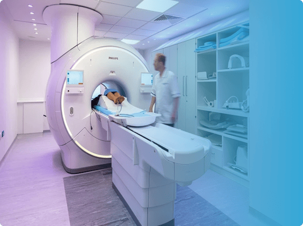
MRI scans rely on the principles of nuclear magnetic resonance (NMR) to generate detailed images of the body's internal structures. The process begins with the patient lying down on a movable table that slides into the MRI machine, which consists of a large cylindrical magnet. This magnet creates a strong magnetic field that aligns the protons in the body's atoms.
Once the protons are aligned, radio waves are emitted into the body, causing the protons to absorb and then release energy. These energy emissions are picked up by the MRI machine's sensors and transformed into detailed images by a computer. The images produced provide a cross-sectional view of the body, allowing healthcare professionals to identify abnormalities and make accurate diagnoses.
MRI scans are particularly effective at visualising soft tissues, such as the brain, spinal cord, and muscles. They can also detect abnormalities in bones and joints. This makes MRI scans an invaluable tool in the diagnosis and monitoring of a wide range of conditions, including tumours, infections, and neurological disorders.
Advantages of MRI scans
One of the major advantages of MRI scans is that they do not expose patients to ionising radiation. This makes them a safer option compared to X-rays and CT scans, which can have harmful effects, especially with repeated exposure. MRI scans are particularly beneficial for pregnant women and young children who are more sensitive to radiation.
Moreover, MRI scans provide excellent contrast resolution, allowing healthcare professionals to differentiate between different types of tissues. This is especially useful when examining complex structures or subtle abnormalities. MRI scans are also highly versatile and can be used to visualise various parts of the body, including the brain, spine, joints, and abdomen.
Additionally, MRI scans can be enhanced with the use of contrast agents, which help highlight specific areas of interest. These agents are injected into the patient's bloodstream and can help healthcare professionals identify tumours, blood vessel abnormalities, and areas of inflammation.
Types of MRI scans
There are different types of MRI machines, each with its specific applications. The most common type is the closed MRI machine, which consists of a cylindrical magnet that the patient enters. Closed MRI machines provide high-quality images and are suitable for most diagnostic purposes. However, some patients may experience claustrophobia or discomfort due to the enclosed space.
To address this issue, open MRI machines have been developed. These machines have a more open design, providing a more spacious environment for the patient. Open MRI machines are particularly useful for patients who are unable to tolerate the confined space of closed MRI machines, such as those with obesity or anxiety disorders.
Another type of MRI machine is the extremity MRI, which is specifically designed to image the extremities, such as the hand, wrist, foot, or ankle. These machines are smaller and more specialised, allowing for more detailed imaging of the targeted area.
Preparing for an MRI scan
Before undergoing an MRI scan, there are certain preparations that need to be made. It is important to inform the healthcare provider about any metal implants or devices in the body, as these can interfere with the magnetic field and affect the quality of the images. Some examples of metal implants include pacemakers, cochlear implants, and certain types of joint replacements.
Additionally, patients may be asked to remove any metallic objects, such as jewellery or watches, as these can also interfere with the magnetic field. Patients should also wear loose, comfortable clothing without any metal fastenings. In some cases, patients may need to change into a hospital gown to ensure that no metal objects are present during the scan.
It is crucial to follow any fasting instructions provided by the healthcare provider, particularly if the MRI scan involves the abdomen or pelvis. This is because certain types of MRI scans require an empty stomach to improve image quality. Patients should also inform their healthcare provider about any allergies or medical conditions they have, as well as any medications they are taking.
What to expect during an MRI scan
During an MRI scan, the patient will be positioned on a movable table, which will slide into the MRI machine. It is important to lie still during the scan to avoid blurring the images. The MRI machine will produce loud knocking or buzzing noises as it operates. To minimise discomfort, patients may be provided with earplugs or headphones to listen to music.
The duration of an MRI scan can vary depending on the specific procedure being performed. Some scans may be completed in as little as 15 minutes, while others may take up to an hour or more. It is important to communicate with the healthcare provider if any discomfort or anxiety arises during the scan.
In certain cases, the healthcare provider may administer a contrast agent through an intravenous line to improve the visibility of certain tissues or structures. This is a routine procedure and is generally well-tolerated. After the scan is complete, patients can usually resume their normal activities unless otherwise instructed by the healthcare provider.
Common uses of MRI scans
MRI scans have a wide range of applications in the medical field. One of the most common uses is in the evaluation of neurological conditions, such as brain tumours, strokes, and multiple sclerosis. MRI scans can provide detailed images of the brain and spinal cord, allowing healthcare professionals to assess the extent of damage or abnormalities.
MRI scans are also valuable in the assessment of musculoskeletal conditions, such as joint injuries, sports-related injuries, and degenerative diseases. By visualising the bones, cartilage, and soft tissues, MRI scans can help determine the severity of the injury and guide treatment decisions. They are particularly useful in orthopaedics and sports medicine.
Additionally, MRI scans are utilised in the detection and staging of various types of cancer. By visualising tumours and their surrounding tissues, MRI scans can help determine the size, location, and extent of cancerous growth. This information is crucial in developing treatment plans and monitoring the effectiveness of treatment.
Risks and safety considerations of MRI scans
While MRI scans are generally considered safe, there are certain risks and safety considerations to be aware of. The strong magnetic field of the MRI machine can cause certain metallic objects to move or become dislodged, leading to potential injury. It is important to inform the healthcare provider about any metal implants or devices in the body before the scan.
Patients with claustrophobia or anxiety may experience discomfort during an MRI scan, particularly in closed MRI machines. Open MRI machines can provide a more comfortable experience for these individuals. In some cases, sedation may be used to help alleviate anxiety and ensure a successful scan.
Patients who are pregnant or may be pregnant should inform their healthcare provider before undergoing an MRI scan. While there is no evidence to suggest that MRI scans are harmful to the foetus, it is generally recommended to avoid unnecessary exposure to strong magnetic fields, particularly during the first trimester.
Alternative imaging techniques to MRI scans
Although MRI scans are highly effective in many cases, there are alternative imaging techniques that may be used depending on the specific situation. X-rays, for example, are often used for imaging bones and detecting fractures. X-rays are quick and readily available, making them a useful initial screening tool.
CT scans, which use X-ray technology along with computer processing, provide detailed cross-sectional images of the body. CT scans are particularly useful for imaging the chest, abdomen, and pelvis. They are often used in emergency situations due to their speed and ability to detect internal bleeding or injuries.
Ultrasound imaging uses high-frequency sound waves to create images of the body's internal structures. Ultrasound is commonly used for imaging the abdomen, pelvis, and developing foetus. It is a non-invasive and safe imaging technique, making it suitable for individuals who cannot undergo MRI or CT scans.
Conclusion
MRI scans are a crucial tool in modern medicine, allowing healthcare professionals to visualise and diagnose a wide range of conditions. By harnessing the power of magnetic fields and radio waves, MRI scans provide detailed images of the body's internal structures without the use of ionising radiation. They offer excellent contrast resolution and can be enhanced with contrast agents for even greater diagnostic accuracy.
Understanding how MRI scans work, preparing for the procedure, and knowing what to expect can help alleviate any anxiety or concerns. While MRI scans are generally safe, it is important to follow the healthcare provider's instructions and inform them about any metal implants or devices in the body.
In conclusion, demystifying MRI scans help us appreciate the remarkable technology behind this diagnostic tool. From their advantages and various types to their common uses and safety considerations, MRI scans continue to revolutionise the field of medicine and contribute to improved patient care. So, the next time you encounter an MRI scan, you'll have a deeper understanding of the science and technology working behind the scenes.

Patient Friendly MRI Scan at UME Health
Our open MRI machine provides a more spacious and less enclosed environment, which can ease any feelings of anxiety or discomfort during the scan. We also allow a friend or family member to sit in the room with you during the scan, if that would make you more comfortable.
UME Group LLP. Registration number: OC333533. A Company Registered in England and Wales. Registered office: 17 Harley Street, London, W1G 9QH ©Copyright 2024 - UME Group LLP. Built and maintained by Pulse Digital Health


